Widespread implementation of screening mammography over the last three decades has resulted in a more than 500% increase in the detection of DCIS. Currently, nearly all DCIS cases are treated equally with surgery and radiation. Foreknowledge of indolent DCIS would guide the selection of patients for active surveillance, thereby avoiding unnecessary morbidity and costs associated with upfront surgery and radiation.
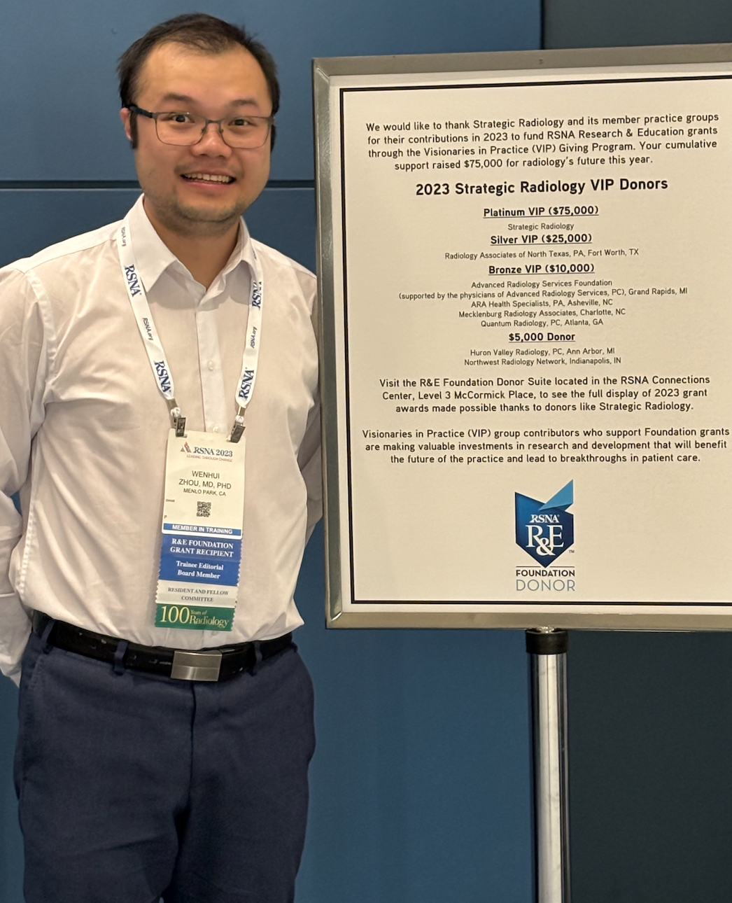
This year, the SR-RSNA Research & Education (R&E) Foundation Resident Seed Grant will help finance the research of Wenhui Zhou, MD, PhD, a 4th year radiology resident at Stanford University Medical School with a strong interest in cancer imaging and interventions. Dr. Zhou’s grant is the fifth in a series of 20 grants to be funded through contributions by individual Strategic Radiology member practices to the RSNA R & E Foundation.
Dr. Zhou recently responded to a series of questions about himself and his current research.
Q: Tell us a bit about yourself and how you became interested in radiological research.
Zhou: During medical school, I was fortunate to have met and worked with supportive mentors who inspired my exploration of imaging research. I was drawn to the new technologies and techniques being developed to visualize, biopsy, and treat cancers. In my residency, I have had the opportunity to further my research interests and collaborate with clinicians and data scientists on imaging projects. It has been a lot of fun, and I am very grateful for the funding support from Strategic Radiology for this academic year.
Q: How big of a problem is the under- and over-diagnosis of DCIS in breast cancer imaging?
Zhou: Widespread implementation of screening mammography over the last three decades has resulted in a more than 500% increase in the detection of DCIS, which is the earliest form of breast cancer and accounts for 20% of all breast cancer diagnosed in the United States. Because of the disease heterogeneity of DCIS, an important imaging challenge for breast cancer diagnosis is to distinguish between indolent and invasive forms of DCIS.
Currently, nearly all DCIS cases are treated equally with surgery and radiation. Foreknowledge of indolent DCIS would guide the selection of patients for active surveillance, thereby avoiding unnecessary morbidity and costs associated with upfront surgery and radiation. Conversely, foreknowledge of invasive DCIS would prompt more extensive treatment upfront to ensure the optimal patient outcome. The goal of our research project is to develop artificial intelligence methods to predict which patients harbor invasive breast cancer occult to current clinical, imaging, and pathology assessment.
Q: What made you think that you could improve the accuracy of risk assessment?
Zhou: Risk assessment of DCIS has been an area of active research. Our approach leverages multiparametric breast MRI and the integration of multimodal data. Breast MRI is the most sensitive modality for DCIS detection, and when incorporated with multiparametric techniques (e.g., high spatiotemporal resolution perfusion and diffusion), it offers complementary anatomical and functional assessment of DCIS.
We plan to develop a computational algorithm to automatically detect and characterize DCIS lesions directly from breast MRI. In addition to quantitative data from MRI, there is non-imaging data from clinical, genomic, and pathology assessment. There is an untapped opportunity to combine these imaging and non-imaging data to predict invasive breast cancer from DCIS. We are optimistic that such an approach will help generate clinically useful risk assessment to guide personalized treatment strategies for DCIS.
Q: How will you pursue your research? Will machine learning play a role?
Zhou: In addition to my clinical and research mentors, I am fortunate to collaborate with a team of graduate students, postdoctoral scholars, and other staff scientists to drive forward this project. We have already curated a well-annotated dataset of patients with preoperative breast MRI, clinical, and histopathologic data. Machine learning is at the centerpiece for aggregating and synthesizing these large datasets for this project. We plan to develop and validate new deep learning methods for breast MRI interpretation and multimodal data integration.
Q: Who will you enlist at Stanford to help? Will this be multidisciplinary research?
Zhou: At the institution level, Stanford offers excellent clinical and research infrastructure, a collaborative environment, and all the elements needed for the success of this project. I will leverage unique resources and potential collaborations within the Center for Artificial Intelligence in Medicine & Imaging (AIMI), the Radiological Sciences Laboratory (RSL), the Integrative Biomedical Imaging Informatics at Stanford (IBIIS), and the Stanford Cancer Institute.
The unique multidisciplinary team supporting this research project is composed of members with complementary expertise in medical imaging analysis, artificial intelligence, and multiparametric breast MRI. I believe that this collaborative team is well-positioned to aggregate and synthesize imaging-based and multidisciplinary biomarkers to improve DCIS diagnostics.
Q: What do you hope to accomplish in this first (and maybe second and third) stage of the project?
Zhou: In the first stage of this project, we aim to develop and validate a deep-learning model to automate DCIS detection and characterization directly from multiparametric breast MRI. This will not only help generate quantitative data useful for downstream applications (e.g., prediction of invasive cancer in DCIS) but also has the potential to standardize the imaging assessment of DCIS.
The second stage of this project is to develop and validate a predictive nomogram using multimodal data from preoperative assessment, for predicting invasive breast cancer in DCIS. The end product will be a user-friendly, clinically actionable, point-of-care tool for radiologists, surgeons, and oncologists to estimate the likelihood of invasive breast cancer for individual patients diagnosed with DCIS.
Hub is the monthly newsletter published for the membership of Strategic Radiology practices. It includes coalition and practice news as well as news and commentary of interest to radiology professionals.
If you want to know more about Strategic Radiology, you are invited to subscribe to our monthly newsletter. Your email will not be shared.